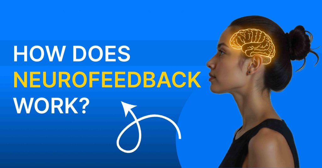

Introduction: What Is qEEG and Why It Matters
qEEG brain mapping is a non-invasive way to measure and visualize how different areas of your brain are working in real time. It records your brainwaves through sensors placed on the scalp and turns them into color‑coded maps that highlight which regions are overactive, underactive, or not working together efficiently.
Unlike a general mental health questionnaire, qEEG (quantitative electroencephalography) looks directly at electrical activity in your brain so treatment can be based on objective data, not just symptoms. This is especially valuable when dealing with complex issues like ADHD, anxiety, depression, sleep problems, or cognitive changes where symptoms often overlap.
qEEG brain mapping matters because it helps answer a question most people never get to ask: “What is my brain actually doing when I feel this way?” With that insight, your care team can create more targeted plans using tools like neurofeedback, medication, or therapy—rather than relying on trial and error.
qEEG vs Traditional EEG, MRI and CT Scan: Key Differences
Most people have heard of EEG, MRI, or CT scans, but are not sure where qEEG fits in. The simplest way to think about it is:
– CT and MRI mainly show **structure**.
– qEEG and EEG show **function**.
Traditional EEG (electroencephalogram) records your brain’s electrical activity and displays it as waveforms, mainly to detect seizures or major abnormalities. It is widely used in neurology to diagnose conditions like epilepsy or to monitor brain activity in intensive care settings.
qEEG uses the same raw EEG data but goes further by applying advanced mathematical and statistical analysis to quantify brainwave activity and compare it to large normative databases. This process converts the waves into numerical values and visual brain maps, highlighting how your patterns differ from what is expected for your age and sex. Where a routine EEG might tell you that activity is “normal” or “abnormal,” qEEG can show which frequency bands (delta, theta, alpha, beta) are too high or too low in specific parts of your brain and how well those regions are communicating with each other. That extra layer of quantitative detail makes qEEG especially helpful for mental health, attention, learning, and performance issues.
MRI (Magnetic Resonance Imaging) and CT (Computed Tomography) scans focus on the **physiological and structural aspects** of the brain. They show the shape, size, and physical integrity of brain tissue—helping detect tumors, bleeding, strokes, structural injuries, and other visible changes. These scans are excellent for ruling out serious medical problems but they do not show how active or inactive different brain networks are during everyday functioning.
qEEG, on the other hand, focuses on the **functional aspects** of the brain—how neural circuits are actually working moment to moment. It looks at brainwave patterns, timing, and connectivity rather than anatomy. This means:
– You might have a completely normal MRI or CT scan (no structural damage), but still have significant issues with attention, mood, sleep, or anxiety.
– qEEG can reveal functional dysregulation—like excessive fast activity linked to anxiety or excessive slow activity linked to inattention—even when structural scans appear normal.
In short:
– CT/MRI = “What does your brain look like physically?”
– qEEG = “How is your brain functioning electrically and network‑wise right now?”
They are not competitors but complementary tools. Structural scans are critical for ruling out medical emergencies and serious disease, while qEEG helps tailor brain‑based interventions like neurofeedback by showing how your brain is functioning.
How qEEG Works: The Technical Process (Explained Simply)
A qEEG brain mapping test usually follows four main steps.
1) Data acquisition (recording)
Small sensors (electrodes) are placed on your scalp using a cap or individual leads, often following a standardized system that covers 19 or more sites. These electrodes pick up tiny voltage changes produced by groups of neurons firing in your brain.
2) Signal processing (cleaning the data)
The recorded EEG signals are digitized and cleaned to remove noise from eye movements, blinking, muscle tension, or electrical interference. This ensures that the analysis is based on true brain activity rather than artifacts.
3) Quantitative analysis (turning waves into numbers)
Specialized software analyzes the cleaned EEG using mathematical methods to calculate power in different frequency bands (delta, theta, alpha, beta, gamma) at each electrode site. It also assesses connectivity—how strongly different areas of your brain are linked in terms of timing and frequency.
4) Comparison to normative databases (creating the brain map)
Your brainwave values are compared against age‑matched, healthy reference databases to calculate how many standard deviations you are from typical patterns (often expressed as Z‑scores). The software then generates color‑coded maps that make it easy to see where activity is higher or lower than expected or where connections are too weak or too strong.
The end result is a set of images and metrics that provide a structured, quantitative picture of your brain’s functional organization. That functional information is what makes qEEG particularly useful for guiding personalized neurofeedback and other brain‑based treatments.
What a qEEG Brain Map Shows
A qEEG brain map gives several key types of information.
1) Power in different frequency bands
– Delta (0.5–4 Hz) – deep sleep, healing, or possible brain injury if elevated while awake.
– Theta (4–8 Hz) – drowsiness, creativity, or excessive daydreaming and inattention when too high.
– Alpha (8–12 Hz) – relaxed wakefulness; too little may indicate difficulty relaxing; too much in some areas can be linked to low drive.
– Beta (13–30 Hz) – alertness and focus; excessive fast beta can relate to anxiety, while low beta can reflect poor concentration.
2) Topographic distribution (where activity is happening)
Brain maps display how these bands are distributed across frontal, central, temporal, parietal, and occipital regions. For example, excessive slow waves in the frontal lobes might be associated with attention and planning difficulties, while abnormal patterns in temporal regions may relate to mood or language issues.
3) Connectivity and coherence
qEEG can measure how synchronized different brain regions are with each other in specific frequency bands. Too much coherence can mean rigid networks and reduced flexibility, while too little may indicate poor communication between regions, affecting functions like working memory, emotional regulation, or processing speed.
4) Deviation from norms (Z‑scores)
Many qEEG systems present deviations from the normative database as Z‑scores, where values above a certain threshold (for example ±2 standard deviations) suggest significant dysregulation. This helps clinicians quickly identify which areas are likely contributing to symptoms.
The map does not read your thoughts or give a diagnosis by itself, but it does reveal functional patterns that can support clinical assessment and guide targeted interventions.
Conditions qEEG Brain Mapping Can Help Detect Patterns For
qEEG is not a standalone diagnostic tool, but it can highlight brainwave patterns associated with many mental health and neurological conditions when interpreted in context by trained clinicians.
Common areas where qEEG is used include:
Some children and adults with ADHD display elevated slow‑wave activity relative to faster beta activity in frontal and central areas. This pattern can correlate with inattention, mental fog, and difficulty sustaining focus.
Excess fast beta activity, especially in certain frontal or central regions, can correspond with chronic worry, restlessness, hypervigilance, and difficulty relaxing. qEEG may also show over‑activation of networks involved in threat detection and arousal.
Some depressed individuals show asymmetries between left and right frontal regions or altered alpha and beta activity that align with low motivation, slowed thinking, and negative mood. qEEG can help identify these patterns and monitor change over time.
– Sleep disorders and insomnia
When the brain has trouble shifting into restful states, qEEG may show persistent fast activity or reduced slow‑wave patterns, even when a person is trying to relax. This can support tailored neurofeedback protocols aimed at calming hyperarousal.
– Autism spectrum conditions
Certain autism profiles have been associated with atypical connectivity and abnormal patterns in specific frequency bands related to sensory processing, social cognition, and communication.
– Traumatic brain injury and cognitive issues
qEEG can reveal areas with slowed activity, disorganized patterns, or connectivity disruptions after concussion or more significant brain injury. This may relate to memory problems, slowed processing, irritability, or fatigue.
– Other conditions
Research continues to explore qEEG’s role in conditions like OCD, PTSD, learning difficulties, and dementia. In each case, qEEG findings must be interpreted with clinical judgment and not treated as a diagnostic label by themselves.
The qEEG Procedure: Step‑by‑Step What to Expect
Most people find qEEG brain mapping straightforward and comfortable. A typical appointment might look like this.
1) Before your appointment
You are usually asked to arrive with clean, dry hair (no heavy oils or styling products) and to avoid caffeine or substances that significantly alter brain activity, if your clinician advises it.
2) Check‑in and history
The clinician reviews your concerns, medical history, sleep, medications, and any previous assessments so they can interpret your brain map in the right context.
3) Electrode cap or sensor placement
A fitted cap with built‑in sensors may be placed on your head, or individual electrodes are applied with conductive gel at standardized positions. You sit comfortably in a chair while the clinician ensures good contact and minimal signal noise.
4) Recording with eyes open and closed
You are asked to sit still, relax, and sometimes look at a point on a screen for a few minutes while your brain activity is recorded with eyes open and then closed. In some protocols, you may be given simple tasks to see how your brain responds under mild cognitive load.
5) Completion and cleanup
After enough clean data is collected, the cap or electrodes are removed and any remaining gel is cleaned from your scalp. The recording portion usually takes 15–30 minutes, though the entire visit may last around an hour including preparation and discussion.
6) Analysis and report preparation
You typically do not receive a full interpretation on the spot; the raw EEG is processed and analyzed with specialized software after your visit. The clinician later reviews the maps, metrics, and your clinical history to prepare a report and treatment recommendations.
7) Feedback session
In a separate appointment, your clinician walks you through the results, explains what the maps show, relates them to your symptoms, and discusses next steps such as neurofeedback, therapy, medical evaluation, or lifestyle strategies.
Safety: Is qEEG Safe and Does It Hurt?
qEEG is considered a safe, non‑invasive procedure because the electrodes only record electrical activity produced by your brain; they do not deliver electricity into the brain. The sensors simply pick up naturally occurring voltage changes at the scalp surface, similar to a very sensitive microphone listening to brain signals.
Most people feel no pain at all during the test—just the sensation of the cap or electrodes on the scalp and possibly some cool gel. There is no radiation, no injections, and no need for sedation. qEEG has been used safely with children, adolescents, adults, and older adults in both clinical and research settings.
Occasionally, people may feel mild discomfort from sitting still for too long, or slight skin irritation if they have sensitive skin where the sensors were attached, but these issues are usually minor and temporary. As with any clinical procedure, trained professionals follow guidelines for preparation, hygiene, and data quality to keep the process both safe and effective.
qEEG Cost and Insurance Coverage
The cost of a qEEG brain mapping assessment can vary widely depending on region, provider expertise, the length of the protocol, and whether it is bundled with other services. In many clinics, qEEG is priced as a specialized assessment rather than a routine test.
In some markets, a qEEG assessment may range from the equivalent of a few hundred to several hundred US dollars, while comprehensive evaluations with additional cognitive testing or imaging may cost more. Some centers package qEEG with a series of neurofeedback sessions or other interventions, offering a combined program rate.
Insurance coverage is mixed: certain policies may reimburse qEEG when it is ordered for defined neurological indications, while others may classify it as investigational or as an out‑of‑pocket service, especially when used primarily for mental health or performance optimization. Because of this, many clinics review your situation individually, check benefits if possible, and give a written estimate before you commit.
If you are considering qEEG, it is wise to ask the clinic:
– What is the exact cost of the assessment?
– Does the fee include the follow‑up interpretation session?
– Will they help you submit claims if your insurance might cover it?
– Are there payment plans if qEEG is part of a larger treatment program?
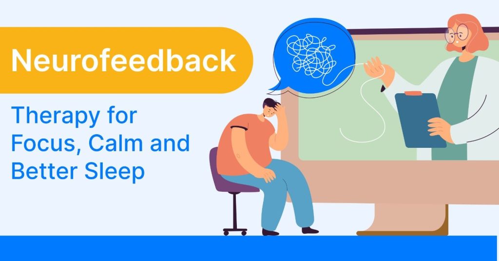
How qEEG Results Guide Neurofeedback Treatment
One of the most powerful uses of qEEG is designing targeted neurofeedback training plans based on your unique brain patterns. Neurofeedback is a form of real‑time EEG‑based training in which you receive visual or auditory feedback whenever your brain activity moves toward a desired pattern, helping you learn to self‑regulate over time.
qEEG results help clinicians decide:
– Which areas of the brain to train – for example, frontal regions for attention and impulse control, or sensorimotor areas for calming the nervous system.
– Which frequencies to reward or inhibit – such as increasing mid‑range beta for sustained focus or reducing excessive high beta that correlates with anxiety.
– How to structure the training plan – including session length, frequency, and progression as the brain changes.
Because qEEG highlights both local dysregulation and network‑level issues, it supports the creation of individualized protocols rather than one‑size‑fits‑all training. Over multiple sessions, many people report improvements in areas like focus, mood stability, sleep quality, stress tolerance, and cognitive clarity as their brain learns more balanced patterns.
The same qEEG framework can also be used to monitor progress by comparing pre‑ and post‑training maps, adding an objective dimension to the subjective improvements you notice.
Common Myths About qEEG Debunked
Because qEEG brain mapping sounds technical, it is easy for myths and misunderstandings to spread. Here are some of the most common myths and why they are inaccurate.
Myth 1: “qEEG can read my thoughts.”
qEEG measures the rhythm and coordination of brain activity, not the content of your thoughts. It can show whether certain areas are overactive or underactive, but it cannot decode specific memories, words, or private ideas.
Myth 2: “qEEG gives a diagnosis by itself.”
qEEG is a tool that shows functional patterns; diagnosis is made by clinicians integrating your history, symptoms, and other assessments. Brain maps support decision‑making but do not independently label you with a condition.
Myth 3: “All abnormal patterns mean disease.”
Brains are individual, and not every deviation from the norm is harmful. Some differences may reflect natural variability, learned skills, or temporary states like fatigue. Interpretation always considers context.
Myth 4: “Once I have a brain map, my brain is fixed that way.”
Brain activity is dynamic and can change with sleep, stress, medication, learning, and targeted interventions. qEEG captures a snapshot in time; follow‑up assessments can document positive changes after training or treatment.
Myth 5: “qEEG is experimental and unsafe.”
EEG has been used for decades, and qEEG builds on this by applying digital analysis. The recording process is non‑invasive and routinely used in clinical and research environments when performed by trained professionals.